|
All Global Research articles can be read in 51 languages by activating the “Translate Website” drop down menu on the top banner of our home page (Desktop version).
Visit and follow us on Instagram at @crg_globalresearch.
First published on July 8, 2021
***
Below are excerpts of the Spanish Team’s research report on Graphene Oxide with links to the full document.
Background
Mr. Ricardo Delgado Martin requests PROVISION OF RESEARCH SERVICES to the UAL named: “DETECTION OF GRAPHENE IN AQUEOUS SUSPENSION SAMPLE”
- On 06/10/2021 1 vial was received by courier, labeled with the following text:
- “COMIRNATY™ .Sterile concentrate. COVID-19 mRNA. 6 doses after dilution.
- Discard date / time: PAA165994.LOT / EXP: EY3014 08/2021 ”
- Origin and traceability: unknown
- State of conservation: refrigerated
- Maintenance during the study: refrigerated
- Coding of the problem sample to be analyzed: RD1
Preliminary observations of the test sample RD1
Description:
- Sealed vial, with rubber and aluminum cap intact, of 2 ml capacity, containing a 0.45 ml cloudy aqueous suspension.
- RNA extraction and quantification is performed
- Presence of uncharacterized nanometric microbiology is observed, visible at 600X in optical microscope

Click screenshot above to access full document in English
Sample processing
1. Dilution in 0.9% sterile physiological saline (0.45 ml + 1.2 ml)
2. Polarity fractionation: 1.2 ml hexane + 120 ul of RD1 sample
3. Extraction of hydrophilic phase
4. Extraction and quantification of RNA in the sample
5. Electron and optical microscopy of aqueous phase
Preliminary analysis: extraction and quantification of Rna in the sample
1. RNA extraction: Kit https://www.fishersci.es/shop/products/ambion-purelink-rna-mini-kit-7/10307963
2. Quantification of total UV absorbance in spectrophotometer NanoDrop™ https://www.thermofisher.com/order/catalog/product/ND-2000#/ND-2000
3. Specific quantification of Rna by fluorescence QUBIT2.0: https://www.thermofisher.com/es/es/home/references/newsletters-and-journals/bioprobes-journalof-cell-biology-applications/bioprobes-issues-2011/bioprobes-64-april-2011/the-qubit-2-0 fluorometer-april- 2011.html
UV absorption spectrum of the aqueous phase of the RD1 sample (Nanodrop team)
Maximum absorption of SAMPLE RD1 (260-270 nm)
- RNA. It presents usual maximums at 260 nm. Total concentration estimated by QUBIT2.0 fluorometry: 6 ng / ul
- The spectrum reveals the presence of a high quantity of substances or substances other than Rna with maximum absorption in the same region, with a total estimated at 747 ng / ul (uncalibrated estimate)
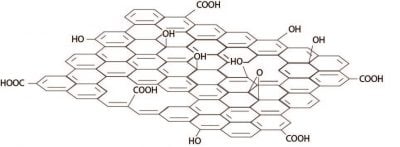
- Reduced graphene oxide (RGO) has absorption maxima at 270 nm, compatible with the spectrum obtained (Thema et al, 2013. Journal of Chemistry ID 150536)
- The maximum absorption obtained DOES NOT ALLOW TO DISCARD the presence of graphene in the sample. The minimum amount of RNA detected by QUBIT2.0 only explains a residual percentage of the total UV absorption of the sample.
OBJECTIVE: Microscopic identification of graphene derivatives
METHODOLOGY:
1. Imaging in optical and electron microscopy
2. Comparison with literature images and reduced graphene oxide standard sample
TRANSMISSION ELECTRON MICROSCOPY (TEM)
Electron microscope JEM-2100Plus
Voltage: 200 kV
Resolution 0.14 nm
Magnification up to x1,200,000
TRANSMISSION ELECTRON MICROSCOPY (TEM)
Electron microscopy (TEM) is commonly used to image graphene nanomaterials. It has become a fairly standard and easy to use instrument that is capable of imaging individual layered graphene sheets.
Conclusions and Recommendations
1. Microscopic study of the sample provides strong evidence for the probable presence of graphene derivatives, although microscopy does not provide conclusive evidence. The definitive identification of graphene, oxidized graphene (GO) or reduced oxidized graphene (rGO) in the RD1 sample requires the STRUCTURAL CHARACTERIZATION through the analysis of specific spectral standard sample comparable to those published in literature and those obtained from the standard sample, obtained with spectroscopic techniques such as XPS, EDS, NMR, FTIR or Raman, among others.
2. The analyzes in this report correspond to ONE SINGLE SAMPLE, limited in total volume available for processing. It is therefore necessary to carry out a significant sampling of similar vials to draw conclusions that can be generalized to comparable samples, recording origin, traceability and quality control during storage and transport prior to analysis.
Read the full report here.
*
Note to readers: Please click the share buttons above or below. Follow us on Instagram, @crg_globalresearch. Forward this article to your email lists. Crosspost on your blog site, internet forums. etc.
|